
Pain is confusing. In fact, it can be so confusing that the source of pain can be a result from a whole other body part or tissue. We call this referred pain, or pain that is perceived at a location other than the source causing the pain. Research shows substantial evidence that pain may refer to distal or proximal structures of the musculoskeletal system. It’s those confusing moments when you see a physical therapist and find out that your hip or back are the actual sources of your leg pain.
It can also be unsettling when you have pain and your health care provider doesn’t want to immediately get an image. Why is this when things can hurt so bad? As providers, when we talk about ordering an MRI, x-ray, CT scan, etc., it should be because there is a strong suspicion that the patient is not appropriate for conservative management after a thorough exam. Conservative management would be any type of therapy or non-invasive procedure. A perfect example of this is with low back pain. Most low back pain typically improves significantly within four weeks and usually does not require any imaging. But, people will often want an MRI to satisfy fears because back pain can rightfully be very scary and debilitating – especially if the patient hasn’t been properly educated on best management.
But then a patient gets an image and another baffling moment takes place. He or she gets referred to physical therapy and the therapist wants to talk about strength, some manual therapy to get joints moving, surrounding tissues impact on pain, and doesn’t even mention anything about the anatomical changes on the image. Now, why is this?!
Well, I’m about to state some known facts in research that may make a lot of people feel less special (which is ultimately a good thing!):
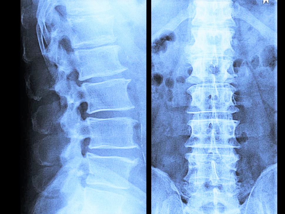
- “Disc protrusions, bulges, and annular tears are commonly present in subjects without low back pain.”¹
- “The incidence of rotator cuff tears increase with age and are present in 50% of asymptomatic subjects by age 70.”¹
- “Standard sequence MRIs performed on volunteers with no history of foot or ankle injury or symptoms revealed anatomic variants commonly associated with peroneal tendon disorders in symptomatic ankles.”¹
What do all these facts mean? Well, disc protrusions, degenerative disc disorders, certain muscle or tendon disorders, etc. are a lot of times just normal wear and tear bodies go through. We probably all have something in our body that is not anatomically perfect…and you know what, THAT IS OKAY! These normal aging processes are happening and usually we don’t have any pain that goes along with it. Meaning, just because an MRI shows a “disc protrusion”, the pain source is usually a combination of things versus just the anatomical change seen on an image. Hence, why a person may end up in physical therapy to help manage all these sources of pain. Which, there is a lot of solid evidence that conservative management like physical therapy is extremely helpful (more on this in another blog post).
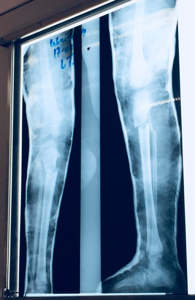
This x-ray is a case where imaging was completely necessary! This poor kiddo needed a surgical consult ASAP (before conservative care of physical therapy) after having a pretty bad leg break. This was one of my patient’s in Tanzania, Africa, that I treated for 3 months.
The bottom line question I always ask my patients when they immediately want an image is: “What would the image provide you?” If it’s peace of mind, I’d challenge a patient to remember the following: In most situations where red flags are not present, a thorough exam has been done, and a patient has not yet had conservative management (cough, cough, physical therapy), an MRI is simply not needed1. It may not change treatment or management of the condition and it’s an expensive addition to monthly bills.
Now, there are absolutely times when an image is completely necessary (see picture). But remember, it’s typically when next steps are being evaluated beyond conservative management. I’m sorry if this post pointed out the inevitable that our bodies are constantly aging, but, I wish healthcare choices could be more straightforward for all of us healthcare consumers out there. Imaging is an area that can add even more frustration to this already confusing world. Ignorance can be bliss with these “wrinkles” that are just part of the human body’s normal aging process.

If you have questions on this post, don’t hesitate to reach out directly!
Email:
Instagram: @dpterin_motion
References:
¹Deyle, G. The role of MRI in musculoskeletal practice: a clinical perspective. Journal of Manual and Manipulative Therapy 2011;19(3): 152-161.
²Flynn, T. W., Smith, B., & Chou, R. Appropriate Use of Diagnostic Imaging in Low Back Pain: A Reminder That Unnecessary Imaging May Do as Much Harm as Good. Journal of Orthopaedic & Sports Physical Therapy 2011;41(11): 838-846.
³Jensen M.C., Brant-Zawadzki M.N., Obuchowski N., Modic M.T., Malkasian D., Ross J.S. Magnetic resonance imaging of the lumbar spine in people without back pain. New England Journal of Medicine 1994;331:69–73.